 (rollover to compare with normal)
 |
|
What Are Its Effects?
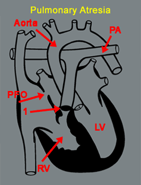
In the normal heart, oxygen-poor blood from the body's tissues is pumped by the right ventricle (RV) through the pulmonary artery (PA) to the lungs for reoxygenation. In Pulmonary Atresia, the route from the right ventricle to the lungs is blocked (1 in diagram).
This situation would prove fatal soon after birth if not for two remnants of the fetal circulatory system that allow some blood from the body tissues to be pumped to the lungs. These are the Patent Ductus Arteriosus (PDA), a small blood vessel that connects the pulmonary artery and aorta and which usually closes soon after birth (not shown in diagram), and the Patent Foramen Ovale (PFO).
The Patent Ductus Arteriosus allows blood to pass from the aorta to the lungs. However, blood returning from the body into the right atrium and right ventricle would be blocked if not for the presence of the small Patent Foramen Ovale (PFO) and, in some cases, a Ventricular Septal Defect. These allow the mixing of blood between the right and left chambers of the heart and permit the passage of blood from the body into the left ventricle (LV), which pumps it to the lungs by way of the aorta and Patent Ductus Arteriosus.
As long as the Patent Ductus Arteriosus, Patent Foramen Ovale, and Ventricular Septal Defect (if present) remain open, the infant can survive, though cyanosis (decreased oxygen in the blood) may develop because less oxygen than normal is carried to the body tissues. Other symptoms that may develop soon after birth include a heart murmur, rapid or labored breathing, lethargy, and irritability.
In cases where the right ventricle is undersized (hypoplastic), the formation of the coronary arteries, which are on the outer surface of the heart and carry oxygen-rich blood to the heart muscle, may be adversely affected. |